Anatomy of the eye - although your eyes are small, they are in fact one of our main senses; vision. Everyone knows that without our eyes we wouldn’t be able to see anything, but often people are unsure of how the eye really works.
So, what is the eye actually made up of?
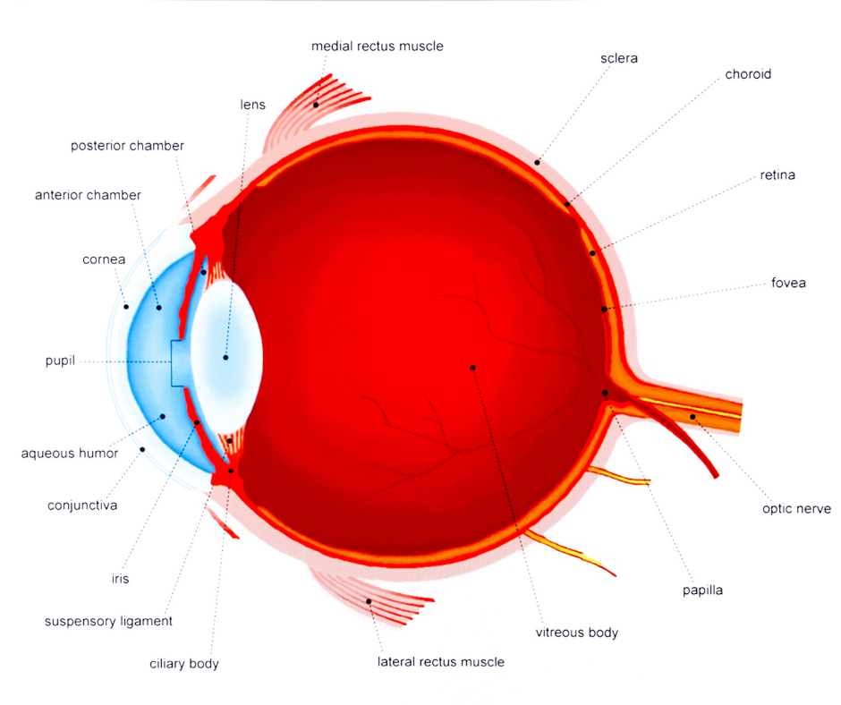
- Iris: the iris is in charge of regulating the amount of light that enters your eye. It also forms the coloured, visible part of your eye in front of the lens.
- Pupil: the pupil is the black circular opening in the centre of the iris. Light passes through this opening into the lens of the eye. The iris is responsible for controlling the amount of light that is let into the eye, this is called dilation and constriction.
- Cornea: the cornea is the transparent circular window of the front of the eyeball. It refracts (changes direction) the light entering the eye onto the lens, which then focuses it onto the retina. The cornea is a very sensitive part of the eye.
- Lens: the lens is a transparent structure that lies behind the pupil. It helps to refract incoming light and focus it onto the retina. People that are affected by cataracts have a cloudy lens.
- Choroid: the choroid lays between retina and the sclera, it absorbs excess light which prevents blurry vision.
- Ciliary body: the ciliary body connects the choroid and the iris.
- Retina: the retina is a light-sensitive layer that lines the inner part of the eye. It is composed of light-sensitive cells known as rods and cones. The retina works in a similar way as a film in a camera.
- Macula: the macula is a yellow spot on the retina which surrounds the fovea.
- Fovea: the fovea forms a small indentation at the centre of the macula and is the area with the greatest concentration of cone cells. When looking at an object, the part of the image that is focused on the fovea is the image registered by the brain.
- Optic disc: the optic disc is the visible portion of the optic nerve. The optic disc identifies the start of the optic nerve where messages from cone and rod cells leave the eye via nerve fibres to the optic section of the brain.
- Optic nerve: the optic nerve leaves the eye at the optic disc and leads to the brain.
- Sclera: the sclera is the white part of the eye, which meets the cornea in the front.
- Rod cells: Rod cells are one of the light-sensitive cells in the retina. There are around 125 million rods, which are responsible for seeing in dim light.
- Cone cells: Cone cells are the second type of light-sensitive cells found in the retina. The human retina contains between 6 and 7 million cones; they function best in bright light and are essential for accurate vision.
(please see anatomy of the eye image)